Introduction
Pectus excavatum (90%) and pectus carinatum (5-7%) are two of the most common type of congenital chest wall abnormalities.
Pectus Excavatum
Pectus excavatum, also known as sunken chest, occurs when the sternum and the ribs grow abnormally, resulting in a concave appearance of the anterior chest wall (Fig. 1). Typically around 4-5 ribs on each side of the sternum are involved, with the degree of asymmetry varying from mild to very severe.
Pectus excavatum is more commonly seen in males (3:1) and occurrence is 1 in 300-400 births. More than 80% of cases are identified within the first 2 years of life, with a more pronounced appearance of the classical clinical features during puberty due to the rapid bone and cartilage growth.
Pectus Carinatum
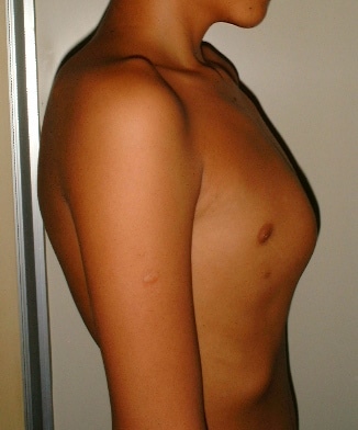
Pectus carinatum, also known as pigeon chest, occurs when the sternum and several ribs protrude anteriorly due to overgrowth of cartilage between the sternum and ribs (Fig. 2).
Pectus carinatum can be classified as either chondrogladiolar, where the middle and lower areas of rib cage arch forward, or chondomanubrial*, where the upper rib cage arch forward (more typically asymmetrical). Pectus carinatum can also be classified by cause, either post-surgical, congenital, or idiopathic.
Pectus carinatum is more common in males and occurs in approximately 1 in 1,500 children. Similarly to pectus excavatum, pectus carinatum becomes more prominent during puberty due to rapid bone and cartilage growth during the growth spurt.
*Chondromanubrial is less common and more difficult to treat due to the affected ribs being shorter and less flexible
Risk Factors
The main risk factors for both pectus excavatum and pectus carinatum are male gender and family history. Patients with Marfan syndrome and Ehlers-Danlos syndrome are also at increased risk of both chest wall disorders
Clinical Features
Patients with pectus excavatum or pectus carinatum are typically asymptomatic and will most commonly present due to cosmetic reasons only.
The majority of patients have symptoms present since birth, with the deformity becomes worse during puberty, however it can also develop post-operatively following thoracic surgery.
In severe cases, the chest wall deformity can lead to cardio-respiratory compromise, with patients presenting with exertional dyspnoea or orthopnoea
Investigations
Unless there is cardiopulmonary compromise, there are no specific diagnostic tests required for pectus excavatum.
For those being considered for operative intervention, CT imaging is required (Fig. 3), especially to aid treatment planning (and assess for any lung compression or mediastinal displacement).
Echocardiography may be indicated in symptomatic patients to assess cardiac function pre-operatively.
Management
Pectus Excavatum
Surgery is the only definitive treatment option for pectus excavatum. Whilst various exercise programmes and external braces are occasionally used as conservative approaches, there is no evidence that they are effective in achieving anatomical correction.
Absolute indications for surgery include clinical features of cardio-respiratory compromise or severe deformity (severity index >3.2*) and severe sternal depression. Any repair should be delayed until the patient is at least aged 8yrs.
The most common surgical approaches are the modified Ravitch procedure or Nuss procedure
*Degree of severity can be determined by the Haller index, which is calculated by dividing the transverse chest diameter by the anteroposterior diameter (on CT scan)
Surgical Management
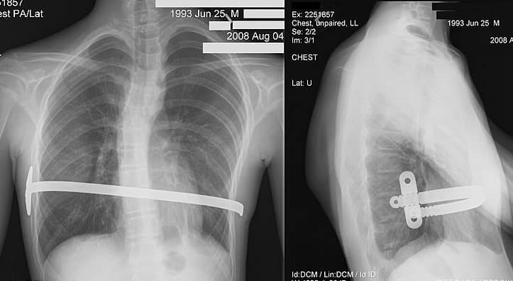
The modified Ravitch procedure involves a submammary incision with subperitoneal excision of the affected costal cartilages, the sternum is elevated via a sternal wedge osteotomy, and a pectus bar is inserted to maintain the elevated position of the sternum (Fig. 4).

Figure 4 – Plain film chest radiograph following a Modified Ravitch procedure
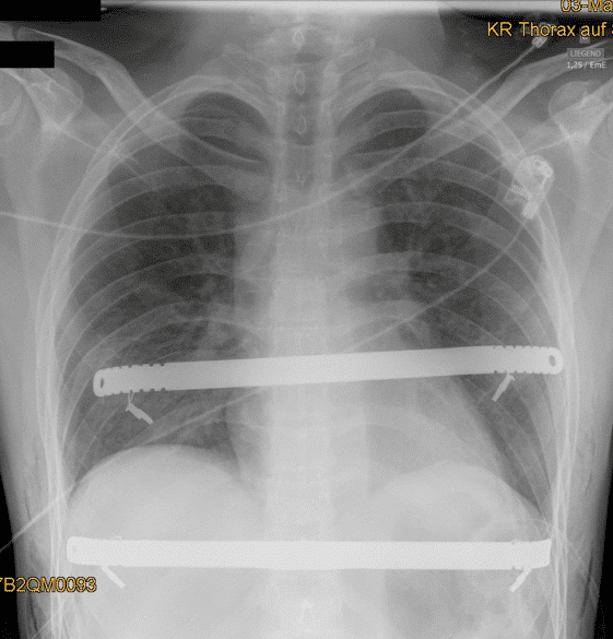
The Nuss procedure involves thoracoscopic placement of a steel bar behind the sternum without excising the costal cartilage, before the bar is rotated to elevate the sternum (Fig. 5).

Figure 5 – Plain film chest radiograph following a Nuss procedure
Pectus Carinatum
Conservative treatment via orthotic bracing is the first line of therapy for pectus carinatum, which is more likely to improve with consistent use because the exogenous compression of the sternum and anterior ribs may over time result in correction. Success rates for conservative treatment are around 65-80%.
If surgical approach is required, this requires a subpericondral resection of the affected costal cartilages, followed by sternal wedge osteotomy for posterior displacement of the sternum and removal of the xiphoid process.
Key Points
- Pectus excavatum and pectus carinatum are two of the most common type of congenital chest wall abnormalities
- The main risk factors are male gender and family history
- Patients are typically asymptomatic and will most commonly present due to cosmetic reasons only, however severe cases can cause cardiorespiratory compromise
- Surgery is the only definitive treatment option for pectus excavatum, whilst conservative treatment via orthotic bracing is usually the first line of therapy for pectus carinatum


