Introduction
Olecranon process fractures are relatively common fractures of the upper limb.
They occur with a bimodal age distribution; occurring in the young following high energy injuries or (more commonly) in older patients following low energy indirect injuries.
In this article, we will look at the pathophysiology, clinical features, investigations and management of olecranon process fractures.
Pathophysiology
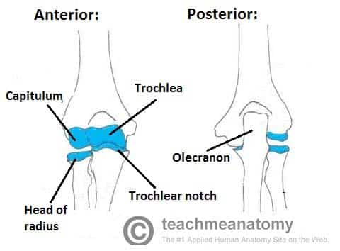
The olecranon is the region of the proximal ulna from its tip to the coronoid process. It articulates with the trochlea of the distal humerus and therefore all olecranon fractures are intra-articular fractures.
The olecranon is the site of insertion for the triceps muscles. Fractures of the olecranon typically result from indirect trauma when a patient falls on an outstretched arm, resulting in the sudden pull of the triceps (and brachialis) muscle. The triceps muscle will also act to further distract the fracture; this is important to appreciate as it influences the management of these injuries.
Less commonly, in younger patients, these are high energy injuries resulting from direct trauma and may be associated with other forearm injuries or fractures.
Clinical Features
Olecranon fractures typically present with a history of falling on an outstretched hand followed by elbow pain, swelling, and lack of mobility.
On examination, there is typically tenderness when palpating over the posterior aspect of the elbow, with a potential palpable defect present. The disruption to the triceps mechanism means often there is an inability to extend the elbow against gravity*. Ensure to check the neurovascular status of the affected limb.
Other injuries associated with a fall on an outstretched hand include wrist ligament and bony injuries and radial head fractures. Therefore, the shoulder and wrist joints should also be examined.
*In minimally displaced olecranon fractures, extension is preserved (albeit tender) due to the soft tissue attachments that remain intact
Investigations
All patients admitted should have routine blood tests taken, including clotting screen and group and save.
Initial imaging should be via plain antero-posterior and lateral radiographs, of both the affected joint and potentially joints above and below too.
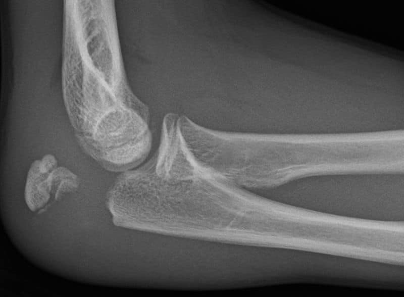
Generally, olecranon fractures are easily identifiable on a lateral projection and with the pull of the triceps have a degree of displacement. There are a variety of different classification systems used in describing olecranon fractures, including the Mayo classification and the Schatzker classification
CT imaging can be useful in evaluating more complex injuries and degree of comminution.
Management
Ensure the patient is resuscitated appropriately and stabilised, prior to definitive management of the fracture. Ensure to provide adequate analgesia.
Treatment is usually guided by the degree of displacement on imaging. Any complex injuries such as fracture dislocations or neurovascular compromise should warrant urgent senior discussion.
Management will often vary between centre, surgical preference, and patient factors, however as an overview:
- Non-operative management is usually indicated for displacement <2mm, with immobilisation in 60-90 degrees elbow flexion and early introduction of range of motion at 1-2 weeks
- There is increasing use of non-operative management for all patients over 75, irrespective of displacement, as whilst the degree of extension may be lost, the functional outcome is often appropriate
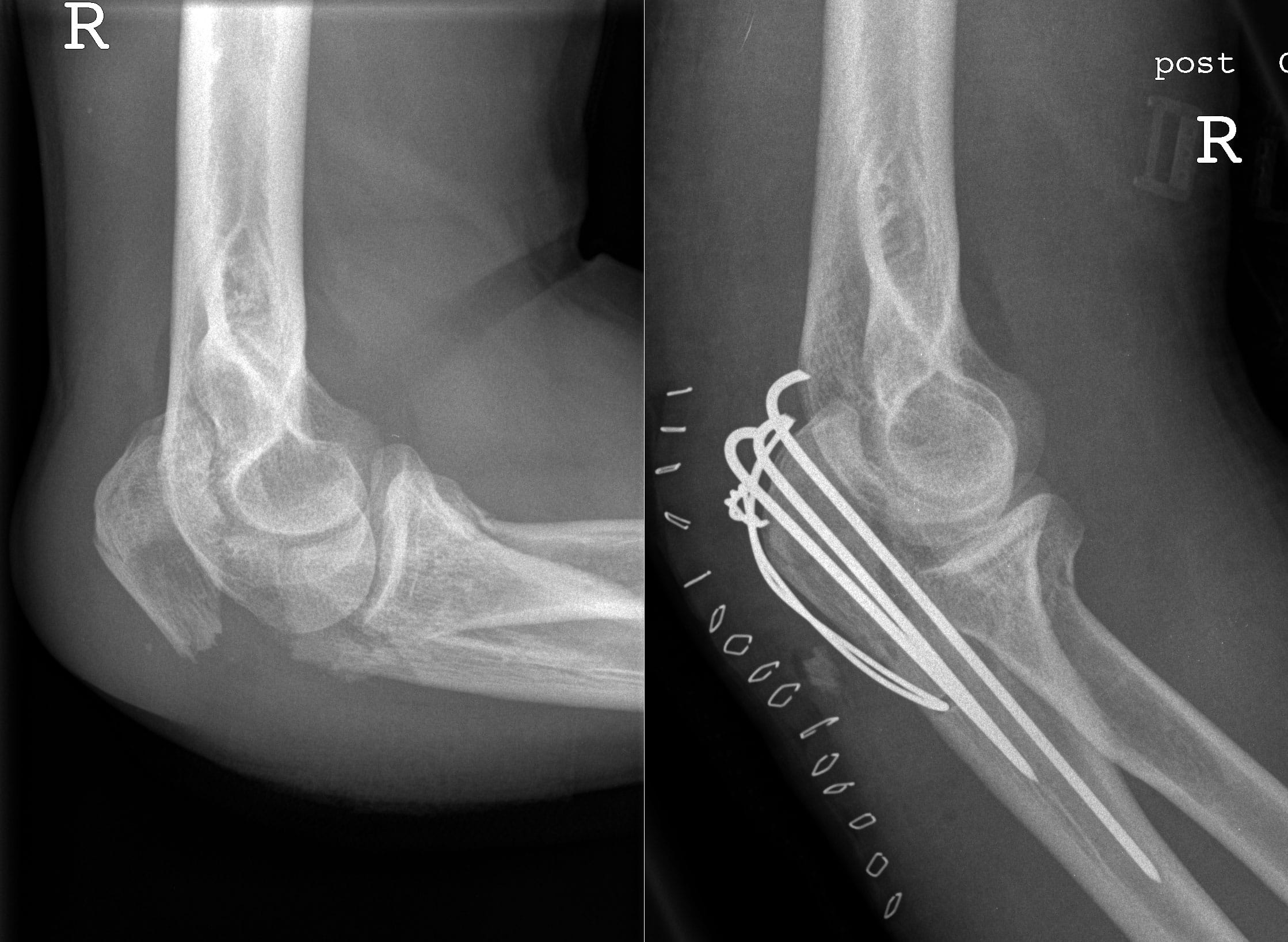
- Operative management is usually indicated for displacement >2mm, requiring (depending on fracture configuration) techniques such as tension band wiring (if fracture proximal to the coranoid process) or olecranon plating (if at level of, or distal to, the coranoid process) may be used
- There is a very high rate of removal of metalwork, as due to the very superficial nature of the injury, it often impacts the patient significantly

Figure 3 – Radiographs of an olecranon process fracture, before and after surgical management
Key Points
- Fractures of the olecranon process are relatively common fractures of the upper limb
- Will often follow falling on an outstretched hand, with tenderness over the posterior aspect of the elbow and a potential palpable defect
- Lateral and antero-posterior radiographs remain the mainstay of initial investigations
- Management can be either operative or non-operative, heavily influenced by degree of displacement


