Introduction
Necrotising fasciitis is a life-threatening rapidly-progressing infection that spreads along the fascial planes and subcutaneous tissue. It has a high mortality, with rates up to 40%, and is a surgical emergency. Fournier’s gangrene is specifically a necrotising infection of the perineum.
The main microbial types of necrotising fasciitis* are:
- Type I is a polymicrobial infection, primarily caused by a mixture of anaerobes (e.g. Bacteroides) and aerobes (e.g. S. aureus); it is the more common of the two subtypes, especially in elderly or co-mordid patients
- Type II is a monomicrobial infection, primarily caused by Streptococcus pyogenes (group A strep), and is more common in healthy individuals with a history of trauma
- It also can be caused by Panton-Valentine leucocidin (PVL)-positive Staphylococcus aureus infections
*Some classifications also include a type III (marine exposure) and a type IV (fungal causes in the immunosuppressed)

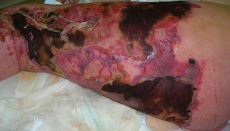
Figure 1 – Necrotising Fasciitis of the left thigh and posterior leg
Gas Gangrene
Gas gangrene is a form of necrotising fasciitis caused by Clostridium species (most commonly C. perfringens), resulting in gas being produced by the bacteria within the tissue.
The clostridial organisms involved produce alpha and beta toxins that lead to extensive tissue damage, alongside producing large volumes of gas within the tissue.
It will present in an equally severe clinical state, however tissue crepitus is often present on light palpation of the affected area. Management is the same as with any necrotising fasciitis.
Risk Factors
The main risk factors for developing necrotising fasciitis include diabetes mellitus, chronic kidney disease, alcohol excess, advanced age or frailty, malnutrition, metastatic cancer, or immunocompromised (e.g. AIDS or recent chemotherapy)
Clinical Features
Whilst some cases may have a recognisable precipitating event of skin breach (e.g. recent trauma, animal bite or scratch, or recent surgery), this is not always the case.
Clinical features will progress rapidly. Patients will complain of severe pain, often out of keeping with the overt clinical signs. Patients will often be haemodynamically unstable and may show signs of multi-organ dysfunction.
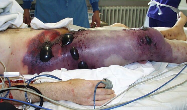
Examination signs are variable, often not representative of the degree or severity of infection present; indeed, overlying skin may appear normal in the early stages. Progression can reveal skin erythema, oedema and signs of skin ischaemia. Late signs include skin crepitus, vesicles / bullae, and obvious skin necrosis.
Differential Diagnosis
Main differentials to consider in suspected cases are cellulitis or myositis, however if there is significant clinical suspicion then urgent exploration should not be delayed.
Investigations
Blood tests will show varying degrees of derangement, certainly with significantly raised WCC and CRP.
A blood gas will likely show a raised lactate +/- metabolic acidosis. There may also be signs of worsening renal function, hyponatraemia, impaired liver function, raised glucose, and coagulopathy as well. Blood cultures should also be taken
Imaging does not have a routine role in the diagnosis of necrotising fasciitis and should never delay management*.
*Plain radiograph in advanced disease may show gas within the tissues and CT imaging can show fluid collections tracking along fascial planes or asymmetrical fascial thickening, including gas within the collections
Risk Scoring
The Laboratory Risk Indicator for Necrotising Fasciitis (LRINEC) score can be used to assist a clinician in the diagnosis of necrotising fasciitis. A score ≤5 is low risk, score 6-7 is intermediate risk, and ≥8 is high risk. This must always be interpreted in the context of clinical suspicion and does not rule out necrotising fasciitis.
It’s important to highlight that Necrotizing Fasciitis is diagnosed clinically, and that any risk score should not postpone surgical intervention, such as debridement. Therefore, it should only be used when the diagnosis isn’t evident.
| Parameter | Range | Score |
| Hb (g/dl) | >13.5 | 0 |
| 11 – 13.5 | 1 | |
| <11 | 2 | |
| White cells (109/L) | <15 | 0 |
| 15 – 25 | 1 | |
| >25 | 2 | |
| Sodium (mmol/L) | <135 | 2 |
| Creatinine (μmol/L) | >141 | 2 |
| Glucose | >10 | 1 |
| C-reactive protein | >150 | 4 |
Table 1 – The Laboratory Risk Indicator for Necrotising Fasciitis (LRINEC) score
Management
Necrotising fasciitis is a surgical emergency and needs immediate resuscitation and debridement if this is deemed appropriate. It is important to remember the high mortality in this disease* and asses each case individually.
Any patient with suspected necrotising fasciitis needs urgent broad spectrum antibiotics (as per local protocol). Resuscitation intravenous fluids should be started and the patient catheterised. Any area of redness or discolouration needs to be marked and time denoted. Early contact to the intensive care team is also required.
Dishwater-like fluid from the wound, or any non-bleeding unhealthy subcutaneous tissue when digitated, fat peeling easily from the fascia, or any unhealthy fascia are all suggestive of the diagnosis.
*In some cases, a palliative approach may be more acceptable to a patient than a disarticulation or proximal amputation
Surgical Management
The only definitive management for necrotising fasciitis is urgent surgical debridement. Intra-operatively, any necrotic tissue should be excised, until only viable bleeding tissue is present.
All cases should be packed following debridement and undergo a relook in 24-48 hours to check for evidence of infection or further necrosis (several repeat debridements may be necessary). Post-operatively, patients should be transferred to a high-dependency or intensive care unit.
Reconstructive surgery might be needed after initial debridement and the infection cleared, using skin grafts or flaps once the infection has been controlled. At this stage, the patient may be appropriate to transfer to a centre with plastic surgery services.
Key Points
- Necrotising fasciitis is a life-threatening rapidly-progressing infection that spreads along the fascial planes and subcutaneous tissue
- Diagnosis is clinical, with patients presenting very unwell and pain out of proportion to clinical signs
- Definitive management is with broad-spectrum antibiotics and urgent surgical debridement

