Introduction
Acute joint swelling is a common presentation to Emergency Departments and can be caused by a wide range of pathologies, a few of which can be life or limb-threatening.
When faced with an acute monoarthritis, the initially assessment is often focussed on differentiating a septic arthritis from the large number of possible differentials (discussed below).
Understanding the differences in the presentation of each pathology is key to aid prompt identification of these conditions.
Clinical Features
Always ensure to establish the onset, site, timeframe of the swelling, any precipitating factors (including trauma or surgery, especially regarding prosthetic joints), and any exacerbating or relieving factors. Clarify the patient’s level of pain and ability to weight bear.
A number of causes of joint swelling will also cause systemic symptoms, therefore asking for any associated fever or rigors, or lethargy is useful. If a rheumatoid cause is suspected, make sure to inspect any other involved joints, and establish if there are any recent gastrointestinal (enteropathic arthritis) or genitourinary symptoms (reactive arthritis), or associated skin changes (psoriatic arthritis).
Previous episodes of the presentation can be an indicator of an acute exacerbation of a chronic issue. Also clarify any past medical history and current medications (including over-the-counter treatments).
Initial Assessment
Any patient who is systemically unwell must be approached in an A to E manner, before further examination of the affected joint can occur.
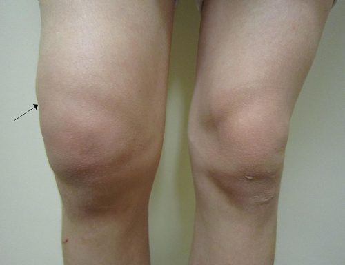
When examining the affected joint(s), ensure to use a look, feel, move framework (see specific examinations). Inspect for any redness, swelling, or skin changes or scars, and make sure to compare this to the contralateral joint.
Check for any focal tenderness and for any evidence of a joint effusion. Range of motion may be affected if the joint is significantly swollen, or if the joint is too painful to move.
Inspect the rest of the body if indicated looking for other joint involvement or any systemic signs that may help elucidate the underlying cause.
Investigations
Investigations performed will depend on the clinical state of the patient.
The majority of patients presenting to the hospital with an acutely swollen limb will have routine bloods, including FBC and CRP to check for infective causes. Addition of an ESR can be useful if a rheumatological cause is suspected, or addition of serum urate if gout is suspected (although can be normal in cases of gout).
Plain film radiographs of the affected joint should also be obtained, especially if any history of trauma, to evaluate for any fracture.
Joint Aspiration
Joint aspiration is the most important investigation when investigating an acute monoarthritis, the technique for this will depend on the joint involved. It is useful to visually inspect the joint fluid for opacity, colour, and presence of frank pus in the syringe.
The aspirate can be sent for white cell count and microscopy (Table 1), culture and sensitivity, as well as light microscopy (for crystals). If a prosthetic joint is affected, joint aspiration should usually be performed in theatres, due to the infection risk.
|
Normal |
Non-Inflammatory Arthritis | Inflammatory Arthritis |
Septic Arthritis |
|
|
Appearance |
Clear |
Clear/Straw-coloured |
Clear or cloudy yellow |
Turbid |
|
White Cells, x106/L |
Normal (<200) |
Moderate (<2000) |
High (>2000) |
Very high (>50,000) |
|
Neutrophils (%) |
Low (<25%) | Low (<25%) | Moderate (<50%) |
High (>75%) |
Table 1 – Synovial Fluid Analysis
Differential Diagnosis
There are a wide range of differentials for patients presenting with an acutely swollen joint.
The main differential to always consider (/exclude) is septic arthritis, however common conditions that present as a swollen joint include haemarthrosis, crystal arthropathies, or rheumatological causes (all discussed below).
Other causes include osteoarthritis, musculoskeletal injury (such as ligamentous or tendon injury, or bursitis), or spondyloarthropathies (such as reactive arthritis, ankylosing spondylitis, or psoriatic arthritis).
Septic arthritis
Septic arthritis is an infection of a joint (discussed further here), most commonly caused by S. aureus. It is important that it is identified and treated quickly as it can cause irreversible articular cartilage damage (leading to severe arthritis) or overwhelming sepsis and even mortality.
Septic arthritis will usually present with a single hot, swollen, painful joint. Patients with septic joints often will not tolerate any passive movement. 60% of septic arthritis patients will be pyrexial, but its absence should not exclude the diagnosis. They will usually have an elevated WCC and CRP.
Joint aspiration is key to diagnosis and the aspirate needs to be sent for Gram stain, leucocyte count, polarising microscopy and culture (Table 1). Ideally aspiration will be performed before any antibiotic administration, however if the patient is unwell then it is important not to delay treatment.
Management is with empirical antibiotics and urgent surgical irrigation and washout. In patients with prosthetic joints a revision surgery will be required.
Crystal Arthropathies
Gout
Gout is an inflammatory arthritis caused by the collection of monosodium urate crystals in a joint. This condition is caused by hyperuricemia leading to crystalisation of the urate in the joint space; importantly however, not all cases of hyperuricemia result in gout (and not all cases of gout will have high serum urate levels).
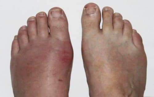
Classically gout affects the 1st MTP joint (Fig. 3), however it can also affect the elbows, knees, wrists, or fingers. Gout is often episodic, with patients experiencing flare ups lasting days or weeks, often brought on by triggers including stress, illness, or dehydration, before a longer symptom-free remission period.
Diagnosis of gout is made by joint aspiration and microscopy, which shows thin, needle shaped monosodium urate crystals in the synovial fluid. Plain film radiographs will often show only a soft tissue swelling, but in recurrent or severe cases “punched out” lesions in the articular bone can be seen.
Acute gout is treated acutely with NSAIDs. Those who experience multiple episodes of gout or with extra-articular features (such as gouty tophi or uric acid nephropathy) can be prescribed prophylactic agents, such as allopurinol, for prevention.

Figure 3 – Gout flare, affecting the left 1st MTP joint
Pseudogout
Pseudogout is an inflammatory arthritis caused by deposits of calcium pyrophosphate crystals within the joint. The condition often mimics gout, however is more likely to affect proximal joints, with the knee and wrist being most commonly affected.
Patients will present acute onset joint swelling, and subcutaneous deposits can also occur near the affected joint. Risk factors for pseudogout include advanced age, hyperparathyroidism, and hypophosphatemia.
Diagnosis of pseudogout is also made by joint aspiration and microscopy, which will show positively birefringent rhomboid-shaped crystals. Pseudo-gout is treated acutely with NSAIDs, as well as treating any underlying cause identified.
Rheumatological
Rheumatoid Arthritis
Rheumatoid arthritis (RA) is an autoimmune disease that can affect any age of patient, although most commonly presents in those aged 40-60yrs. The small joints in the hands and the feet are more commonly affected (often sparing the DIPJs), however it can occur in any joint in the body, being symmetrically affected.
Patients will present with swollen, painful, and red joints, with stiffness that is usually worse in the morning. Patients may also have associated fatigue, lethargy, pyrexia, or weight loss, or other MSK-type presentations, including synovitis, tenosynovitis, rheumatoid nodules, or bursitis.

Figure 4 – Severe Rheumstoid Arthritis affecting the hands, producing a “Swan Neck” deformity in the fingers
Blood tests will show raised inflammatory markers (mainly CRP & ESR) and may also have a normocytic anaemia. Rheumatoid factor (RF) and ACPA levels can also aid a diagnosis of RA. Plain radiographs can show features of RA, with early signs of soft tissue swelling, periarticular osteopenia, and juxta-articular erosions, and late signs of a narrowed joint space, subluxations, and destructive changes.
Treatment will usually be initiated by rheumatologists, and can include NSAIDs for pain, before starting DMARDs (e.g. sulfasalazine or methotrexate) or biologic agents (e.g. infliximab or etanercept).
The EULAR Classification
The diagnostic criteria for RA is the EULAR classification which classifies 4 categories and ≥6/10 indicated a definite RA.
- Joint Distribution (0-5): 1 large joint = 0 points, 2-10 large joints = 1 point, 1-3 small joints = 2 points, 4-10 small joints = 3 points, >10 joints (at least one small joint) 5 points
- Serology (0-3): Negative RF AND negative ACPA = 0 points, low positive RF or low positive ACPA = 2 points, high positive RF or high positive ACPA = 3 points
- Symptom Duration (0-1): <6 weeks = 0 points, ≥6 weeks = 1 point
- Acute Phase Reactants (0-1): Normal CRP and normal ESR = 0 points, abnormal CRP or abnormal ESR = 1 point
Spondyloarthropathies
Spondyloarthropathies are a group of conditions comprising of Psoriatic Arthritis, Ankylosing Spondylitis, Reactive Arthritis, and Enteropathic arthropathy. They are classified as being seronegative conditions (RF negative), and are associated with HLA-B27.
They all can present with “axial arthritis” (those affecting the spinal and SI joints), or affecting any joint in the body as an oligoarthritis or monoarthritis, as well as ability to cause enthesitis and dactylitis.
Most diagnosis for the spondyloarthropathies are made clinically, being confirmed with subsequent biochemical and radiological investigations.
Traumatic
Trauma to a joint will often cause swelling. It is important to be able to differentiate whether a swollen joint following trauma is able to be managed conservatively or whether further intervention will be required.
Haemarthrosis
Bleeding into a joint cavity is referred to as haemarthrosis. The most common cause of haemarthrosis is a traumatic injury, however it can also occur either non-traumatically in patients with bleeding disorders, such as haemophilia, or those on anticoagulants.
In patients with an acutely swollen joint following trauma, haemarthrosis should always be suspected. There may also be a concurrent ligamentous or meniscal injury that has specifically caused the bleeding (e.g. ACL containing a genicular artery).
Initial investigations will include routine bloods, including clotting, and plain film radiographs of the affected joint. A joint aspiration can be performed, especially in patients with large symptomatic haemarthrosis, which can also make a definitive diagnosis.
Initial management includes rest, ice, compression, and elevation of the joint (RICE), and ensuring sufficient analgesia for the pain. The majority of cases are managed conservatively, including the correction of any underlying coagulopathies.
Key Points
- There are a wide range of pathologies that can present with acutely swollen joint
- Ensure to consider septic arthritis in the initial assessment
- Rheumatological causes include gout, rheumatoid arthritis, and spondyloarthropathies
- Most cases of haemarthrosis are managed conservatively


