Introduction
A hernia is defined as the protrusion of part or whole of an organ or tissue through the wall of the cavity that normally contains it.
There are numerous types of abdominal wall hernias, the most common of which are inguinal, femoral, and incisional hernias, however many other types have been described, as outlined below.
Abdominal wall hernia are more likely in those with a high BMI, multiparous, poor nutritional state, male gender, and older age.
Uncomplicated hernia of the abdominal wall can often undergo elective surgery for repair, either as a suture repair or a mesh repair. However, those presenting strangulated or obstructed will often require emergency surgery.
During emergency repairs, the level of contamination (for instance if there is bowel perforation or non-viable bowel requiring resection) will factor into decision making regarding the use of mesh*
*If the risk of subsequent mesh infection is too high, primary repair can be a more appropriate option, even with a large hernia defect
Epigastric Hernia
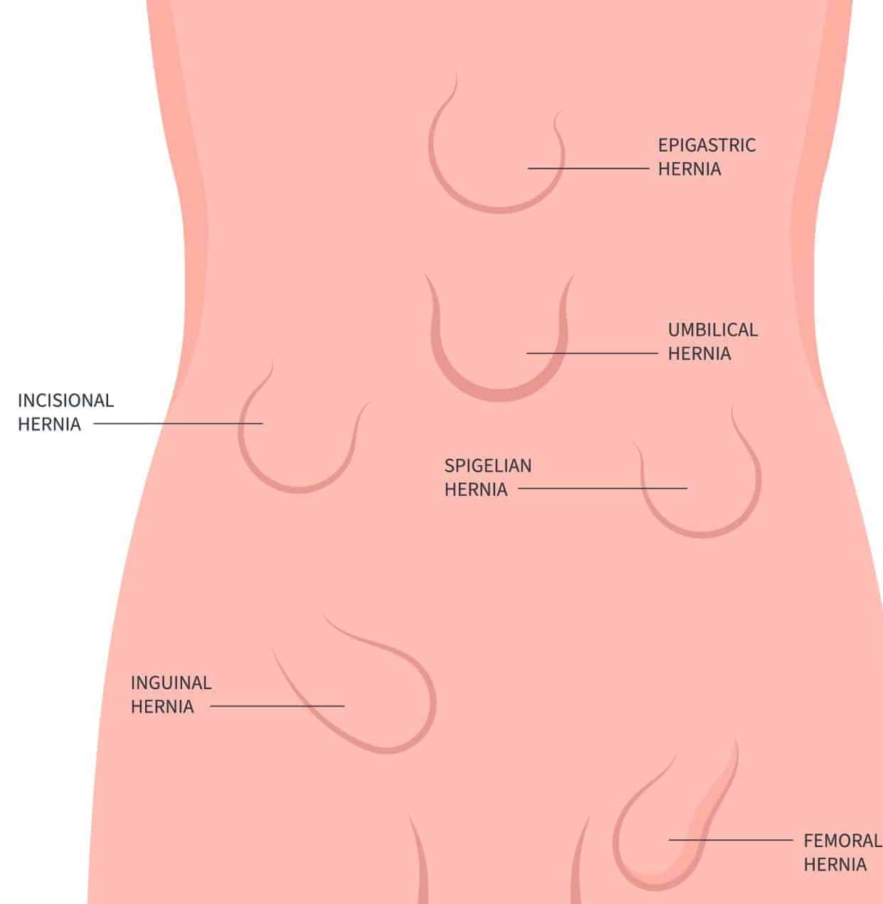
An epigastric hernia is an abdominal wall hernia occurring in the upper abdominal region (above the umbilicus), through the fibres of the linea alba (Fig. 1). Around 1-5% of all abdominal hernia undergoing an operation are epigastric hernia.
Whilst often asymptomatic, they will present as a reducible lump in the epigastric region, however less commonly will present incarcerated or strangulated.
The majority of cases if symptomatic will require surgical repair, which can be done either via an open approach or a laparoscopic approach. A mesh will be used in the repair depending on the size of the hernia defect (approximately a defect >1cm will require a mesh)
Divarification of the Recti
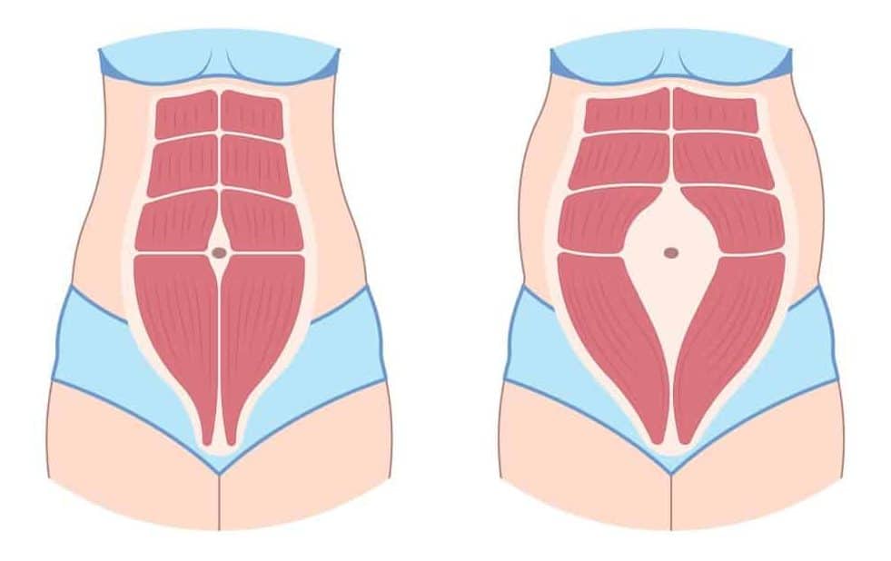
Divarification of the Recti (or rectus diastasis) is a stretching of the linea alba, resulting in a widening of the gap between the rectus abdominus muscles (Fig. 3). As there is no defect in the abdominal wall, it is not a hernia.
Risk factors for its development include advanced age and multiparity. It often presents with a bulge when sitting up, however no discrete lump or mass is present.
As no hernia is present, generally no surgery is required and the mainstay of initial treatment is with physiotherapy to strength the abdominal wall. Any surgical intervention is often cosmetic.
Umbilical and Paraumbilical Hernia
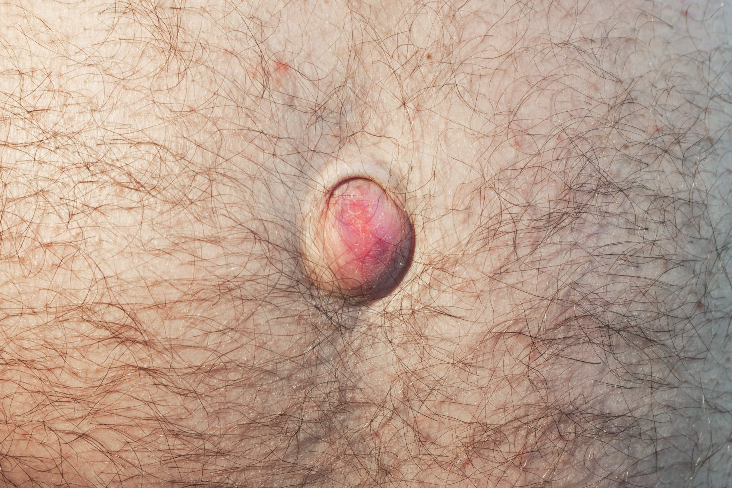
An umbilical hernia is a hernia occurring through a defect in the abdominal wall at the umbilical cicatrix (Fig. 4), whilst a paraumbilical hernia is a hernia occurring through a defect in the abdominal wall adjacent to the umbilicus.
These are common hernia*, many being incidentally identified and patients having had them present for a number of years. Like all abdominal wall hernias, risk factors include obesity and multiparity.
They often only contain pre-peritoneal fat, however will increase in size over time and may eventually contain abdominal viscera. As such, they can present acutely, with either strangulated or obstructed contents.
The majority of cases if symptomatic will require surgical repair, which can be done either via an open approach or a laparoscopic approach. A mesh will be used in the repair depending on the size of the hernia defect (approximately a defect >1cm will require a mesh).
*Newborns can also be affected by congenital abdominal wall defects, either omphalocoele or gastrochschisis
Spigelian Hernia
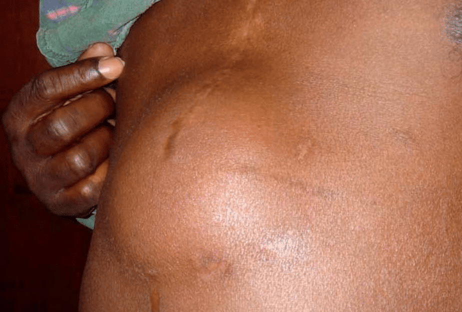
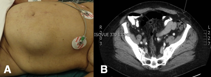
A spigelian hernia is a rare form of abdominal hernia occurring around the level of the arcuate line at the semilunaris (Fig. 5)
Clinically, they present as a small lump at the lower lateral edge of the rectus abdominus muscle. They have been associated with higher rates of cryptorchidism (present in 75% of Spigelian hernias in male infants), likely due to a failure in gubernaculum development.
They have a high risk of strangulation and therefore should be repaired urgently. Either via an open approach or a laparoscopic approach, and a mesh should be placed if feasible.
Obturator Hernia
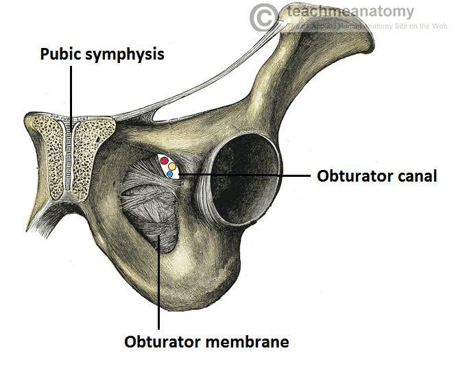
An obturator hernia is a hernia of the pelvic floor, occurring through the obturator foramen, into the obturator canal (Fig. 6). They are more common in women (due to a wider pelvis) and in frailer older patients (due to lack of fat within the obturator canal, leading to a larger space for potential herniation).
Patients will classically present with a lump in the upper medial thigh. Due to their high risk of obstruction, many patients will present either with strangulation or obstruction.
In around half of cases, compression of the obturator nerve passing through the obturator canal will result in a positive Howship-Romberg sign (hip and knee pain exacerbated by thigh extension, medial rotation, and abduction).
Given the high risk of strangulation, obturator hernia should be repaired urgently.
Richter’s Hernia
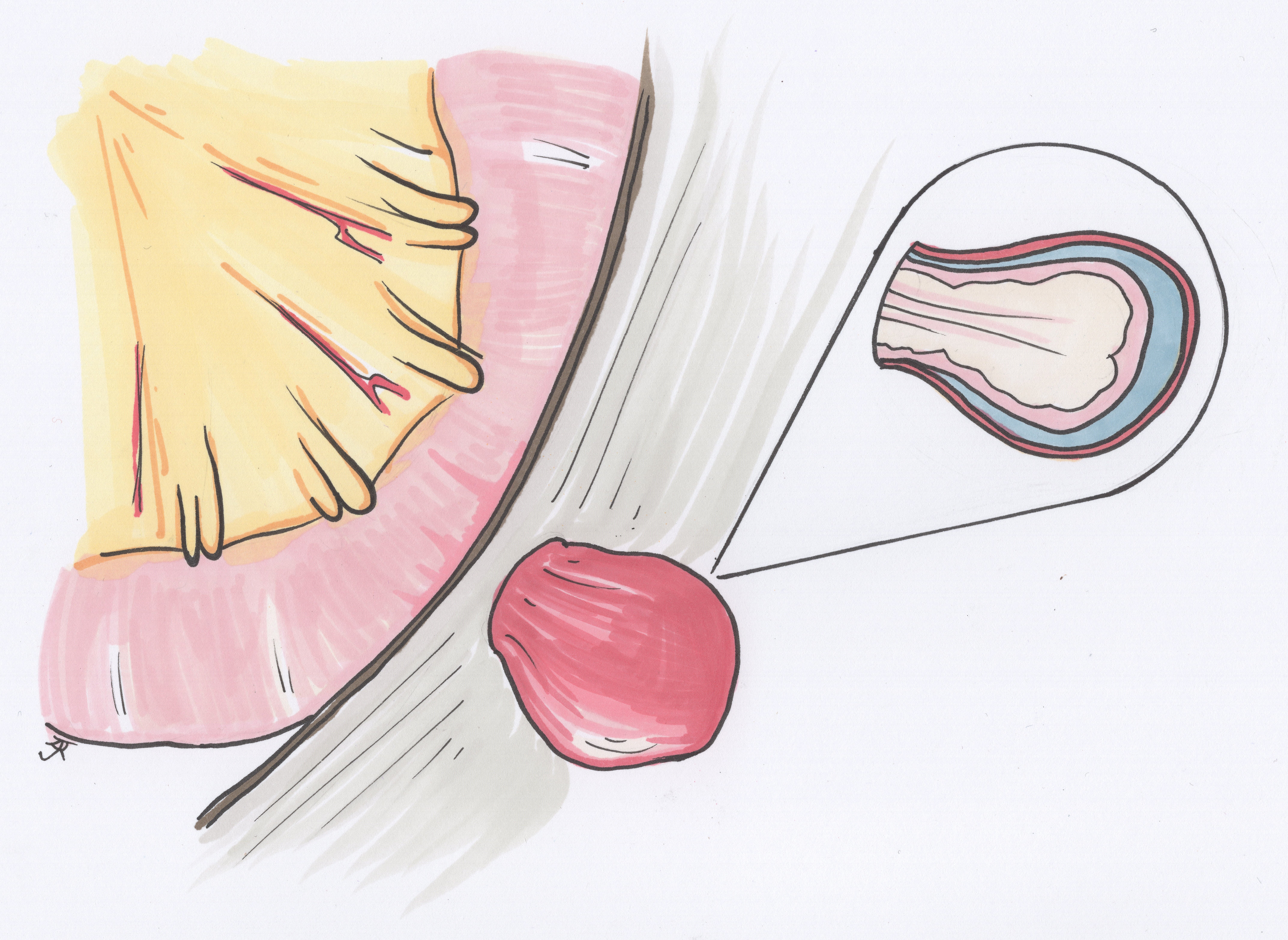
A Richter’s hernia is a partial herniation of bowel (through any hernial orifice), whereby only the anti-mesenteric border becomes involved in the hernia. As such, only part of the bowel lumen is within the hernial sac (Fig. 7), but the compromise to the blood supply to the involved bowel wall means that risk of ischaemia is high.
Patients will present with a tender irreducible mass at the herniating orifice and will have varying levels of obstruction (purely dependent on how much bowel circumference is involved). Importantly, patients may not present with symptoms of obstruction initially.
However due to risk of bowel ischaemia, these are often surgical emergencies that need urgent surgical intervention.
Lumbar Hernia
Lumbar hernias are rare posterior hernias, that typically occur spontaneously or iatrogenic following surgery (classically following open renal surgery). They present as a posterior lump, often with associated back pain.
Other Abdominal Wall Hernias
There are a few rare eponymous abdominal wall hernia that have been recorded in the literature:
- A Littre hernia is any type of abdominal hernia which contains a Meckel’s diverticulum
- An Amyand hernia is a form of inguinal hernia which contains the vermiform appendix (seen in less than 1% of inguinal hernia)
- A De Garengeot hernia is form of femoral hernia which contains the vermiform appendix
Key Points
- Epigastric hernia occur in the upper midline through the fibres of the linea alba
- Umbilical hernia occur through the linea alba around the umbilical region
- Spigelian hernia occur at the semilunar line around the level of the arcuate line
- Obturator hernia occur through the obturator foramen into the obturator canal
- Richter’s hernia are partial herniation of bowel involving the anti-mesenteric border

