Introduction
Around 2% of attendances in the Emergency Department are due to bite injuries, most commonly from dogs, cats, and humans. If left untreated, bites have a high risk of becoming infected.
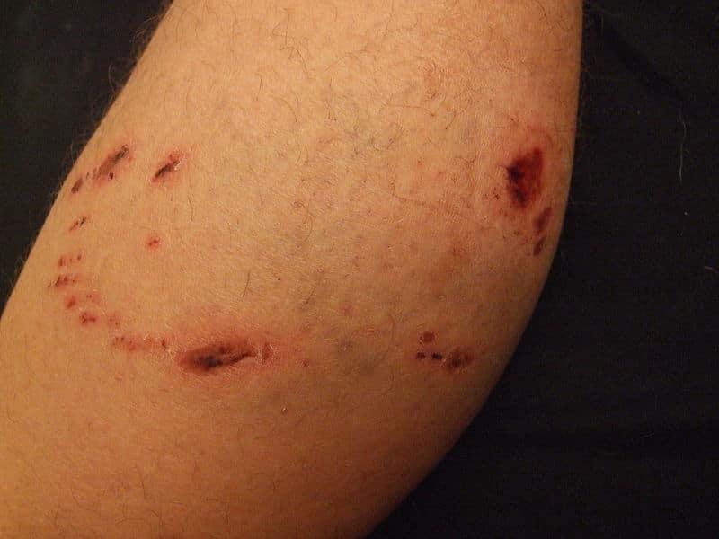
The type of animal involved will determine the specific injury that occurs; cats have a weaker bite but sharper teeth, therefore cause deep puncture wounds, whilst dogs have a strong bite, therefore can also cause crush injuries and lacerations.
Human bites commonly occur as a “fight bite”, whereby a closed fist punches a person’s mouth, however they do subsequently sustain a bite injury. This can occur commonly into the MCP joint (knuckle with closed fist), thus can result in septic arthritis if left untreated.
Pathogens from Bites
Bite wounds can be polymicrobial, as a result of skin flora and organisms from the mouth of the animal*.
Importantly for human bites, certain viruses, such as HBV, HCV, and HIV, can be also be transferred, therefore appropriate prophylaxis may be required.
Certain organisms that are introduced through bites can lead to more systemic illness (Table 1)
*Rabies (caused by the lyssavirus) is not common in the UK but should be considered in other countries as well; rabies vaccine and immunoglobulin may be considered as after symptoms of rabies show, the disease often is fatal
| Organism | Located | Symptoms |
| Pasteurella multocida & Pasteurella cannis | From both cat and dog bites | Early onset (within 12–24hrs) with local cellulitis, purulent discharge, and lymphangitis |
| Staphylococcus aureus & Streptococci | From both dog and human bites | Usually onset after 24 hours |
| Bartonella henselae | Causes “cat scratch disease”; from both cat and flea bites | Occurs in children and immunocompromised individuals; typically is self-limiting |
| Streptobacillicus moniliformis | Causes “rat bite fever”; from rats, squirrels, ferrets, and other rodents | Results in fever, vomiting, headache, myalgia, and a rash, between 3 to 10 days following contact. |
Table 1 – Notable Organisms from Animal Bites
Clinical Features
Bites from animals commonly occur on the dominant arm or leg, where the victim has tried to stop the attack (in children, facial injuries are more commonly present).
Whilst most bites will appear small and uncomplicated superficially, deeper structures may have been affected, therefore it is important to always check the neurovascular status of the affected limb.
For any bites to the hand or foot, ensure to check for any damage to the tendons by assessing the movement of the joint (flexor digitorum superficialis for the proximal interphalangeal joint and flexor digitorum profundus for the distal interphalangeal joint in the hand).
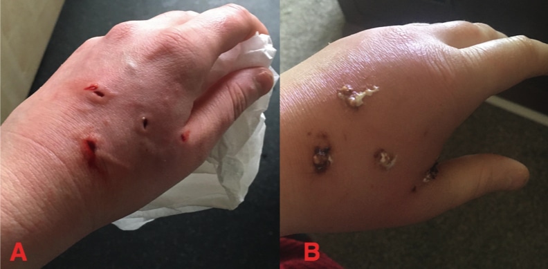
The most important sequela of a bite is infection. Infection can develop within 1-2 days following the bite*, presenting with erythema, discharge, localised pain, or swelling (Fig. 1). Infection can spread rapidly, and can lead to deep space infections (thenar/hypothenar space, space of parona tracking to carpal tunnel) or even necrotising fasciitis. Tracking lymphangitis can often be seen with spreading cellulitis.
*Cat bites have double the infection risk of a dog bite due to the small puncture wounds that introduce bacteria
Investigations
Patients presenting following a bite should obtain a plain film radiograph of the affected region to ensure no residual foreign material is deep within the wound, as well as ensuring no fracture is present as well under the bite.
For those with features of infection, routine bloods (including FBC and CRP) should be taken; any purulent material present should be swabbed. Suspected cases of necrotising fasciitis should be investigated and managed as such.
Management
Initial management of bites should involve preventing further bleeding from the bite and wound cleaning. If there is any ongoing bleeding from the bite, apply continuous pressure and a gauze dressing to the wound. Photographs of the wound should also be taken if possible for documentation.
Tetanus immunisation should be given if a patient has not had a booster within the last 10 years. Antibiotics should be commenced, as per local guidelines. Always consider admission for patients with worsening infection or signs of systemic involvement.
Surgical Management
Small bite wounds should be thoroughly washed with saline. Formal surgical washouts should be considered when bites occur near or into the joint or those with significant associated trauma to soft tissues or mangled limbs.
Any non-viable or damaged tissue should be debrided, with severe injuries returning for a relook after 24-48hrs. Carpal tunnel decompression in the upper limb may be required if tracking pus is seen proximally into the palm and carpal tunnel.
Any joint involvement or associated fracture should necessitate a surgical washout. If done early, metal work may be inserted but usually definitive fixation may be done in 48 hours time after the primary washout. Joint washouts are particularly important in the hands to reduce the risk of infection.
Key Points
- Animal and human bites have increased risk of infection
- Early surgical washout and debridement may allow for primary closure but severe infection is often staged and left to heal by secondary intension.
- Prophylaxis treatment should be given for patients at risk of tetanus or rabies
- Musculoskeletal involvement or neurovascular compromise should be escalated promptly – any involvement of joints and septic arthritis should be admitted for a washout as well.
With thanks to Dr. Charles Yuen Yung Loh MBBS MSc MS MRCS


