Introduction
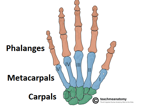
The metacarpal bones form the intermediate component of the skeletal hand, extending between the carpus of the wrist and the phalanges of the fingers.
Fractures of the metacarpals are extremely common and represent a significant proportion of the acute hand surgery workload.
In this article, we shall look at the assessment and management of metacarpal fractures.
Common Fracture Types
There are common fracture patterns observed in metacarpal injuries:
- Boxer’s Fracture – a fracture of the 5th metacarpal neck, which typically occurs from a clenched fist striking a hard surface (such as a wall); these are typically very stable fractures and can be managed with a period of buddy taping and then full mobilisation
- Bennett’s Fracture – an intra-articular fracture of the 1st metacarpal base, with a palmar and ulnar fragment, this fracture pattern typically requires reduction and surgical fixation.
- Rolando Fracture – an intra-articular fracture of the 1st metacarpal base, with a Y or T configuration. This fracture pattern typically requires reduction and surgical fixation.
Clinical Features
Metacarpal fractures usually occur as a result of a traumatic injury to the hand. The clinical features are non-specific, such as pain and swelling in the area overlying the fracture, and reduced range of motion at the affected metacarpophalangeal joint.
In any patient with a hand injury, it is first important to ascertain their hand dominance and occupation, as this may impact decisions regarding treatment.
On examination, look for any wounds suggestive of an open fracture and ensure to check the neurovascular status of the hand. When assessing movement, it is important to check for rotational deformity, extensor lag, and the flexor and extensor tendon function.
Assessing Rotational Deformity
A metacarpal fracture with significant rotational deformity is a common indication for surgical intervention. Rotational deformity is evaluated by asking the patient to make a fist.
In a normal hand, the fingertips will point towards the scaphoid tubercle. In a metacarpal fracture, the finger may be rotated internally or externally, with the fingertip no longer pointing towards the scaphoid tubercle. In severe cases, the fingertips may overlap; this is referred to as “scissoring” of the digits
Investigations
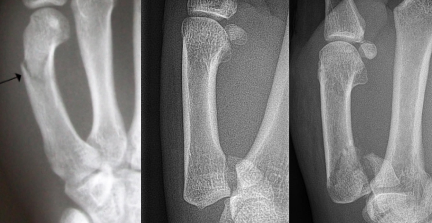
The first line investigation for all suspected metacarpal fractures are plain film radiographs of the hand (Fig. 2), encompassing postero-anterior, oblique, and true lateral views.
CT imaging can be useful in the assessment of fracture-dislocations (Fig. 3) at the carpo-metacarpophalangeal joints, as well as pre-operative planning of complex or comminuted injuries.
Management
Ensure to remove any jewellery from the affected hand and provide adequate pain relief. Elevate the affected hand in a high-arm sling to reduce swelling.
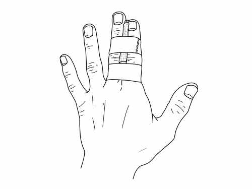
The majority of metacarpal fractures are managed without surgery; the fracture is immobilised with either buddy taping (Fig. 4), ulnar gutter, or volar resting splint. A repeat radiograph is often performed after 1 week of immobilisation to assess for any displacement (which may necessitate surgery).
The fracture is typically immobilised for a total of 3-4 weeks, after which another plain film radiograph is performed to assess for bone healing. The patient may experience stiffness at the immobilised joints, therefore hand therapy rehabilitation is often required after the period of immobilisation.
Surgical Management
There are few absolute indications for surgical intervention, the decision to operate is based on patient factors and injury factors.
The relative indications for surgical intervention include rotational deformity, intra-articular involvement, significant volar angulation, significant shortening, or an unstable fracture pattern
There are many operative approaches to metacarpal fixation; broadly, there are two main techniques:
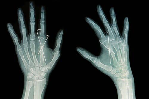
- Closed reduction and percutaneous fixation with K-wires – the fracture is reduced and then held in place with temporary wires, which are removed in 3-4 weeks’ time (Fig. 5)
- Open reduction and internal fixation – an incision is made in the skin and the fracture is reduced under direct vision; metal screws or plates are then used to hold the fracture in the correct place

Figure 5 – Percutaneous K-wire fixation of a metacarpal fracture
Key Points
- Fractures of the metacarpals are extremely common and represent a significant proportion of the acute hand surgery workload
- When assessing a metacarpal fracture, it is important to check for rotational deformity, extensor lag, and the flexor and extensor tendon function
- The first line investigation for all suspected metacarpal fractures are plain film radiographs of the hand
- The majority of metacarpal fractures are managed without surgery, using buddy taping and repeat imaging


