Introduction
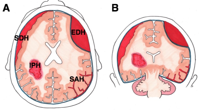
A subdural haematoma (SDH) is a collection of blood that forms in the subdural space, the space between the dura mater and the arachnoid mater (Fig. 1).
They can be classified as acute (< 3 days after injury), subacute (3-21 days), or chronic SDH (>21 days), or as simple (no associated parenchymal injury) versus complicated (associated underlying parenchymal injury)
Pathophysiology
Bleeding in a SDH occurs from tearing of the bridging veins that cross from the cortex to the dural venous sinuses, which are vulnerable to deceleration injury.
This subsequently leads to accumulation of blood between the dura and arachnoid and results in a gradual rise in intracranial pressure (ICP). This can lead to herniation and brainstem death if left untreated.
Risk Factors
The majority of acute SDH occur due to trauma, however can occur in the absence of any trauma (commonly as chronic SDHs).
Risk factors for SDHs include increasing age*, alcohol excess, epileptics (as prone to falls and head injury), and those with clotting disorders or taking anti-coagulants.
*Bridging veins are more vulnerable to tear due to brain atrophy, causing stretching of these veins
Clinical Features
Patients can present with clinical features including altered level of consciousness, headaches, focal neurology, features of raised intracranial pressure (such as blurred vision, worsening headache), or even seizure activity.
Clinical features of an acute SDH occur quickly, whilst those of a chronic SDH have a latent period of weeks (or even months) before symptoms appear. Indeed, the initial injury may be relatively trivial or forgotten, especially in the elderly patients or those with recent alcohol excess.
In children, it is important to survey for other injuries with suspected SDH as there might be signs of non-accidental injury.
Differential Diagnosis
For acute SDH, the main differentials include extradural haematoma, subarachnoid haemorrhage, or intracerebral haemorrhage or infarction.
In chronic SDH, other differentials include space-occupying lesions (such as tumour or abscess), meningitis or encephalitis, or dementia.
Investigations
Patients should have initial routine bloods, including FBC, CRP, U&Es, LFTs, and a clotting, which will also aid in assessing for differential diagnosis. A group and screen will be requiring for any surgical intervention required.
The gold-standard initial imaging modality* for a suspected SDH is a non-contrast CT scan of the head (Fig. 3). SDH should show a crescent-shaped collection of blood over one hemisphere, with or without associated midline shift.
*A skull plain film radiograph may be used in non-accidental injury work up in paediatrics
Temporal Changes on CT Imaging
Subdural haematomas will appear differently on CT imaging, depending on the timing of presentation:
- Acute – diffusely hyperdense
- Subacute – heterogenously hyperdense or isodense
- Chronic SDH – diffusely hypodense
An acute-on-chronic subdural haematoma will present with areas of hyperdensity within a hypodense haematoma.
Management
In an acute setting, patients should be approached initially in a systematic A to E assessment. Any concerns of raised ICP should also be managed appropriately.
All patients on anticoagulation should have this reversed appropriately, which may require discussion with haematology colleagues. Patients are often also started on anti-epileptic medications for 1 week after presentation of a SDH.
For those that present with a SDH following a fall, they should also be investigated for potential underlying reasons for falls. This can often require a multidisciplinary approach, with input from geriatricians, physiotherapists, and occupational therapists as required.

Figure 3 – A left sided frontal parietal subdural haematoma (arrow) with associated midline shift
Definitive Management
Management should be based on the size and clinical features, and indeed not all cases require surgery. Conservative management is generally appropriate for small acute SDH that do not cause significant midline shift or cisternal encroachment without any significant neurologic impairment.
When surgical intervention is required, for an acute SDH, a trauma craniotomy flap is often performed, whereby a large opening in the skull is created to evacuate the haematoma and relieve the associated mass effect. For large acute SDHs, there is often a significant mass effect present, therefore the bone flap is often left out at surgery, termed a decompressive craniectomy; for smaller acute SDH, it may be appropriate to replace the bone flap at the end of the operation.
For chronic SDH, surgical intervention can be either a burr hole craniotomy with irrigation or a twist-drill craniotomy with drain placement. Using a drain has been shown to decrease recurrence rates and mortality without increasing complications.
Complications
Complications following a SDH include cerebral oedema and raised ICP, seizures, herniation, persistent vegetative state, and permanent neurological or cognitive deficits. There is also an increased risk of recurrent haematoma formation following an initial SDH.
Key Points
- A subdural haematoma (SDH) is a collection of blood that forms between the dura mater and the arachnoid mater
- The majority of acute SDH occur due to trauma
- Patients can present with altered level of consciousness, headaches, focal neurology, or features of raised ICP
- Gold standard initial investigation is via non-contrast head CT scan


