Introduction
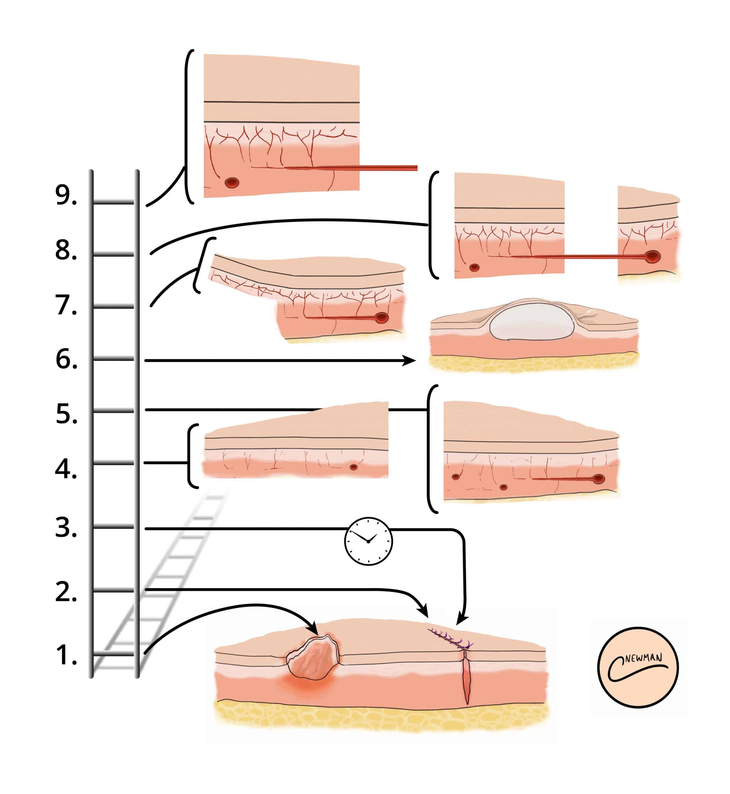
Skin grafts and skin flaps are two surgical techniques that are commonly utilised by plastic surgeons when a defect cannot be closed by primary or secondary intention (Fig. 1, see Reconstructive Ladder).
The key differences between a graft and a flap is in regards to its blood supply; a skin graft receives its blood supply from the recipient site though the vascular bed, whilst a skin flap brings its blood supply from the flap donor site.
They can be used to cover sizeable defects however contra-indications to their use include infection, known skin cancer, and certain patient co-morbidities (immunosuppression, current smoker, poorly controlled diabetes)
Skin Grafts
A skin graft has no blood supply, and therefore depends on the vascularised bed where it is placed. Skin grafts are often utilised as part of the management of extensive skin damage, such as those caused from deep burns, following large skin excision procedures, or poorly healing ulcerating lesions
Several considerations need to be made when choosing a donor site and follow the principle of “replacing like-for-like” when possible; this includes the amount of skin required, the colour and texture of the donor skin, and if hair growth is required at the recipient site.
There are two types of skin graft:
- Split-skin thickness skin graft (SSG) – does not contain the whole dermis
- Full-thickness skin graft (FTSG) – contains the whole dermis (also transplanting hair follicles)
Physiology of Graft Take
Graft take is the incorporation of the new graft into implanted site and can be divided into four main stages:
- Adherence – Fibrin bonds form immediately and is a normal physiological response
- Plasmatic imbibition – occurs around days 2-4 following application of skin grafts, whereby initially fluid (serum) migrates and is absorbed into the skin graft
- Revascularisation – occurs around day 2-3, whereby a vascular network slowly begins to be established; direct anastomosis occurs between vessels from the skin graft and those from the wound bed/ recipient site (inosculation), and then neovascularisation (formation of new blood vessels) follow inosculation
- Remodelling of scar tissue
Skin grafts must heal by developing a new blood supply. For that, the skin graft must be firmly adherent to the recepient wound bed. They can fail (i.e. not obtain an adequate blood supply) for a number of reasons, including haematoma or seroma formation under the graft, infection (commonly Streptococcus spp.), shearing forces, an unsuitable bed (e.g. avascular wound beds, such as tendons or bone), or technical error.
Signs of graft failure include pallor or discolouration at the graft site, skin graft non-adherence to the wound bed, and evidence of localised infection, systemic features (malaise, lethargy), or even full thickness necrosis* (occurs 1-2 weeks after grafting)
All skin grafts undergo contraction, either primary or secondary. Primary contraction relates to the immediate contraction or recoil of freshly harvested skin and is more pronounced in FTSG. Secondary contracture describes the process of contraction once the graft has been applied to its bed and healing is underway and is more pronounced in SSGs.
*Superficial necrosis can be seen in a full thickness graft and may be replaced by healthy tissue

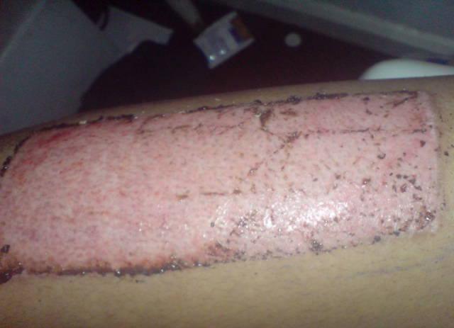
Figure 2 – Donor site 8 days following a split thickness skin graft; note the islands of pigmented epithelium scattered on the donor site which represent healing of the donor site)
Full Thickness Skin Grafts
A full thickness skin graft contains the full thickness of the epidermis and dermis. They are commonly used to cover areas with optimal vascular availability (Table 1), as they have higher energy requirements and are therefore may be more prone to graft failure. Once the graft is harvested, there is no epidermis left behind at the donor site and therefore this site must be closed using sutures (direct closure).
|
Donor site |
Common recipient site(s) |
| Postauricular skin | Face |
| Upper eyelid skin | Contralateral eye defect |
| Supraclavicular skin | Face |
| Flexural skin (e.g. antecubital fossa) | Hand surgery, flexion contractures |
| Thigh and abdominal skin | The palms of the hands or soles of the feet |
Table 1 – Common Donor Sites and Recipient Areas for Full Thickness Skin Graft
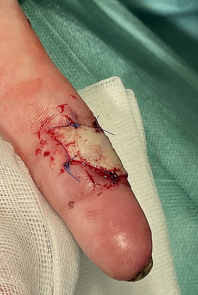
The graft is harvested using a scalpel, taking the epidermis and dermis. All subcutaneous fat is removed, in a process called de-fatting, with tissue scissors, to then be sutured into place at the donor site (Fig. 3)

Figure 3 – A full thickness skin graft from the left forearm on the left index finger
Split Thickness Skin Grafts
A split thickness skin graft contains the full epidermis with a variable thickness of dermis, leaving dermal remnants at the donor site to allow for re-epithelization. Split thickness grafts are commonly used for skin defects that are too large for a full thickness graft. The most commonly used donor site is the thigh, however other donor sites include the forearm, torso, and lower leg.

Figure 4 – Sideview of a dermatome, with the blade (24) removing the epidermis (40) and part of the dermis (38)
Skin grafts are commonly harvested using a dermatome. The dermatome is applied to the skin with downward and forward pressure to harvest the graft (Fig. 4). A dermatome is designed to harvest a graft with consistent dermal thickness from almost any anatomical location on the trunk or limbs*. Other options for harvesting grafts include the oscillating Goulian knife or free hand knives (Watson knife).
*The versatility of this piece of equipment makes it a useful tool for the surgical management of burns patients due to the increased availability of donor sites
Skin Flaps
A skin flap is where tissue is transferred from a donor site to recipient site along with its corresponding blood supply.
Skin flaps are thought to provide better cosmetic results than skin grafting (Fig. 5), as the skin tone and texture are usually better matched. Additionally, they have a reduced chance of failure in comparison to skin grafts.
However, flap failure remains a potential complication of the procedure*. This can occur due to issues with either the arterial supply, presenting with signs of pallor and reduced perfusion, or venous supply, presenting with features of venous congestion.
*Arterial issues need immediate return to theatre, whilst venous congestion often responds to conservative treatment.

Figure 5 – A healing skin flap, used to cover an excision site
Classification
Flaps can be classified via their tissue type, blood supply, or location.
Tissue Type
For tissue types, this is based on the compositions that are utilised in the flap, including cutaneous flap, fasciocutaneous flap, musculocutaneous flap, or muscle flaps
Blood Supply
Based on blood supply, three definitive types of flap are possible:
- Axial flap – a designated subcutaneous artery that runs beneath the flaps longitudinal axis (the artery can be a direct, facsiocutaneous, or musculocutaneous artery)
- Random flap – no designated named artery that provides blood supply to the flap; blood supply via the subdermal plexus
- Pedicled (or perforator) flap – the tissue is completely raised on a named vessel from the donor site and then transferred to the recipient site; this can be as a pedicled flap or free flap

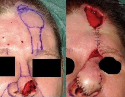
Figure 6 – A nasal reconstruction using a paramedian forehead flap, a type of axial flap
Location Subtypes
When defined by location, flaps can be classified as either local, regional, or free flaps
Local flaps are harvested from a contiguous site and are commonly used for facial defects (Fig. 6), fingertip injuries, or defects on the limb. These can be further classified into:
- Advancement flap – The skin is moved directly forward
- Rotation flap – The skin is rotated around a pivot point to cover an adjacent defect
- Transposition flap – Moves laterally in relation to the pedicle to cover an adjacent defect
Regional (or pedicled) flaps are harvested from the same anatomical region but not directly adjacent. The attached skin (or pedicle) will be tunnelled under the intact tissue, or laid over intact skin forming what is known as a skin bridge, which can then detached from the donor site in a second procedure.
Free (or distant) flaps are harvested from a different anatomical region entirely. The tissue and named fasciocutaneous artery are separated from the donor site before being reattached at the recipient site using microsurgical techniques. Examples of free flaps are seen in Table 2.
| Flap | Donor site |
Vessel |
| Deep inferior epigastric perforator (DIEP) | Skin and subcutaneous tissue of lower abdomen, spares the rectus abdominis | Deep inferior epigastric artery |
| Transverse Rectus Abdominis Myocutaneous (TRAM) | Skin, subcutaneous tissue, and part of the rectus abdominus | Deep inferior epigastric artery |
| Latissimus Dorsi Myocutaneous Flap (LDMF) | Skin, subcutaneous tissue, and part of the latissimus dorsi | Subscapular artery |
| Thoracodorsal artery perforator (TAP) | Skin and subcutaneous tissue of lateral back, sparing the latissimus dorsi | Thoracodorsal artery |
| Anterolateral thigh (ALT) | Skin and subcutaneous tissue of anterolateral thigh (can include vastus lateralis muscle) | Descending branch of lateral circumflex artery |
Table 2 – Specific Examples of Common Free Flaps
Key Points
- Skin grafts and skin flaps are two surgical techniques that are commonly utilised when a defect cannot be closed by primary or secondary intention
- A skin graft has no blood supply and therefore depends on the vascularised bed where it is placed
- A skin flap is where tissue is transferred from a donor site to recipient site along with its corresponding blood supply

