Introduction
- Introduce yourself to the patient
- Wash your hands
- Briefly explain to the patient what the examination involves
- Ask the patient to remove their bottom clothing, exposing the entire lower limb
- Offer a chaperone to the patient
Start by inspection of the patient but be prepared to be instructed to move on quickly to certain sections by the examiner.
Inspection
Ask the patient to stand up, if they are able to do so, and inspect from the back as well as the front.
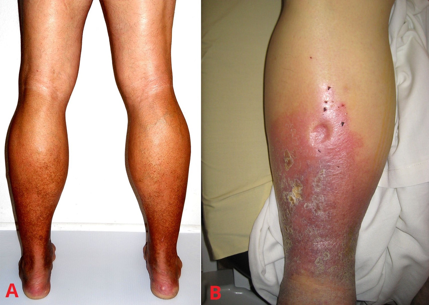
- Look for signs associated with venous insufficiency
- Varicose eczema, haemosiderin staining (brown patches), or atrophie blanche (white patches with dark borders)
- Ulceration, commonly located on the medial aspect around the ankle (gaiter area)
- Lipodermatosclerosis
- Hardening of the skin distally and swelling of the calf
- Look for any varicosities present in the lower limbs, inspecting the course of both the long and short saphenous veins
- Long saphenous vein: medial side of the leg, running anterior to the medial malleolus, posterior to the medial condyle of the knee, before draining into the femoral vein in the medial femoral triangle
- Short saphenous vein: posterior side of the leg, running posterior to the lateral malleolus, along the lateral border of the calcaneal tendon, before draining into the popliteal vein between the two heads of the gastrocnemius
- Scars indicating previous surgery
- Any obvious varicose veins
- Remember that varicose veins can start in the pelvis, causing varicosities in the buttocks and/or vulva
- Look for signs associated with arterial disease (assessing for potential suitability of compression therapy, if required)
- Loss of hair
- Nail thickening
- Surgical scars
- Ulceration
- Particularly checking the pressure points and between the toes
In practice, the examination would normally end at this point as all patients require an ultrasound scan, as described below, prior to any definitive surgical treatment. However the following examinations are good to know for any exam.
Palpation
- Palpate the saphenofemoral junction (4cm lateral and inferior to the pubic tubercle)
- Feel for any saphena varix (large varicosity at sapheno-femoral junction)
- Perform the cough test, asking the patient to cough and feeling for any thrills or dilatations at the junction
Percussion
- Perform the tap test (if any obvious varicosities present) to assess the competency of the valves
- Place one hand at the saphenofemoral junction and the other hand on any varicosity visible
- Tap at the saphenofemoral junction and feel for the percussion with the other hand
- Move distally down the varicosity until unable to feel the percussions
Special Tests
- Perform Trendelenburg’s test, with the patient positioned supine, to assess the level at which the defect is occurring
- Raise the affected leg and massage the leg down in an attempt to drain maximal venous blood from the limb
- With the leg still elevated, place a tourniquet around the thigh
- Ask the patient to stand up and observe for any signs of varicose veins reappearing
- If the varicose veins do not fill back up, this indicates the problem is above the tourniquet level (i.e. at the saphenofemoral junction); if the varicose veins fill back up, this indicates the problem is below the tourniquet level
- Repeat lower down the limb as necessary, assessing for the level where the defect is occurring
- Perform Perthe’s test, with the patient positioned supine, to assess the functioning of the deep veins
- Place the tourniquet around the thigh
- Ask the patient to raise their heels off the ground ten times
- Look for collapse of the superficial veins
- If the superficial veins collapse, indicates deep veins are functioning (as blood is returning through the deep system); if the superficial veins remain dilated, suggests there may be a problem with the deep system
Complete the Examination
Remember, if you have forgotten something important, you can go back and complete this.
To finish the examination, stand back from the patient and state to the examiner that to complete your examination, you would like to perform:
- Doppler Ultrasound Scan, assessing site of venous incompetence (saphenofemoral and saphenopopliteal junction) and the patency of the deep venous system.
- Peripheral Vascular Examination
The primary treatment for varicose veins is compression stockings for symptomatic relief and preventing ulceration. However, compression stockings cannot be used in patients with peripheral vascular disease, as this will worsen their symptoms and cause arterial ulcers, so a peripheral vascular examination with an ABPI are an essential part of the examination.

